
Retinal Disease
Choroidal Melanoma
Choroidal melanoma is a cancer of the eye that develops in the pigmented cells of the choroid. The choroid is composed of layers of blood vessels that nourish the back of the eye, located between the sclera and the retina. It is unusual for a choroidal melanoma to spread to other parts of the body, but similar to skin melanoma, it is possible.
Important to Know
Many cases are found during routine eye examinations.
Most patients exhibit no symptoms of choroidal melanoma and it is only discovered through routine eye examinations.
Retina Comparison
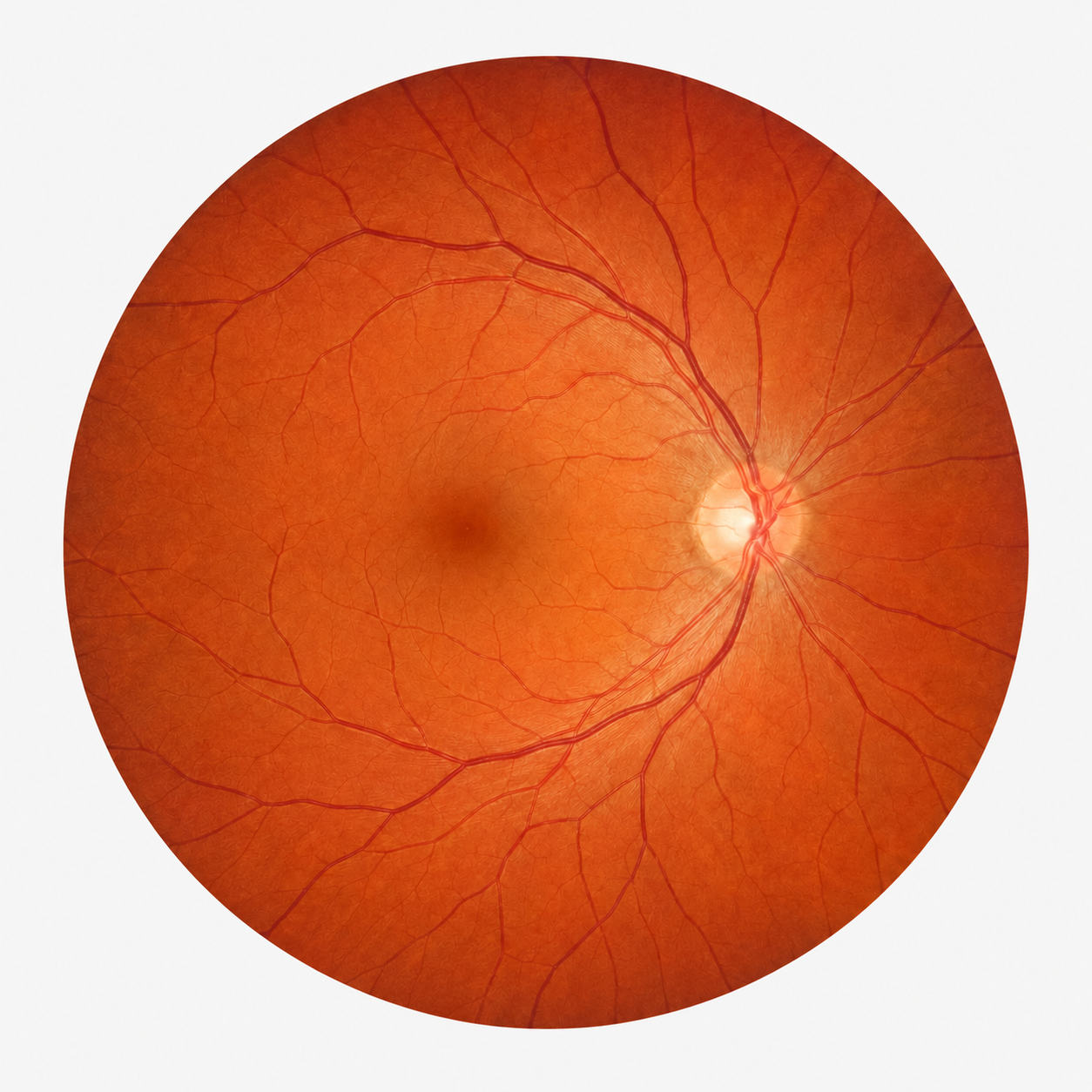
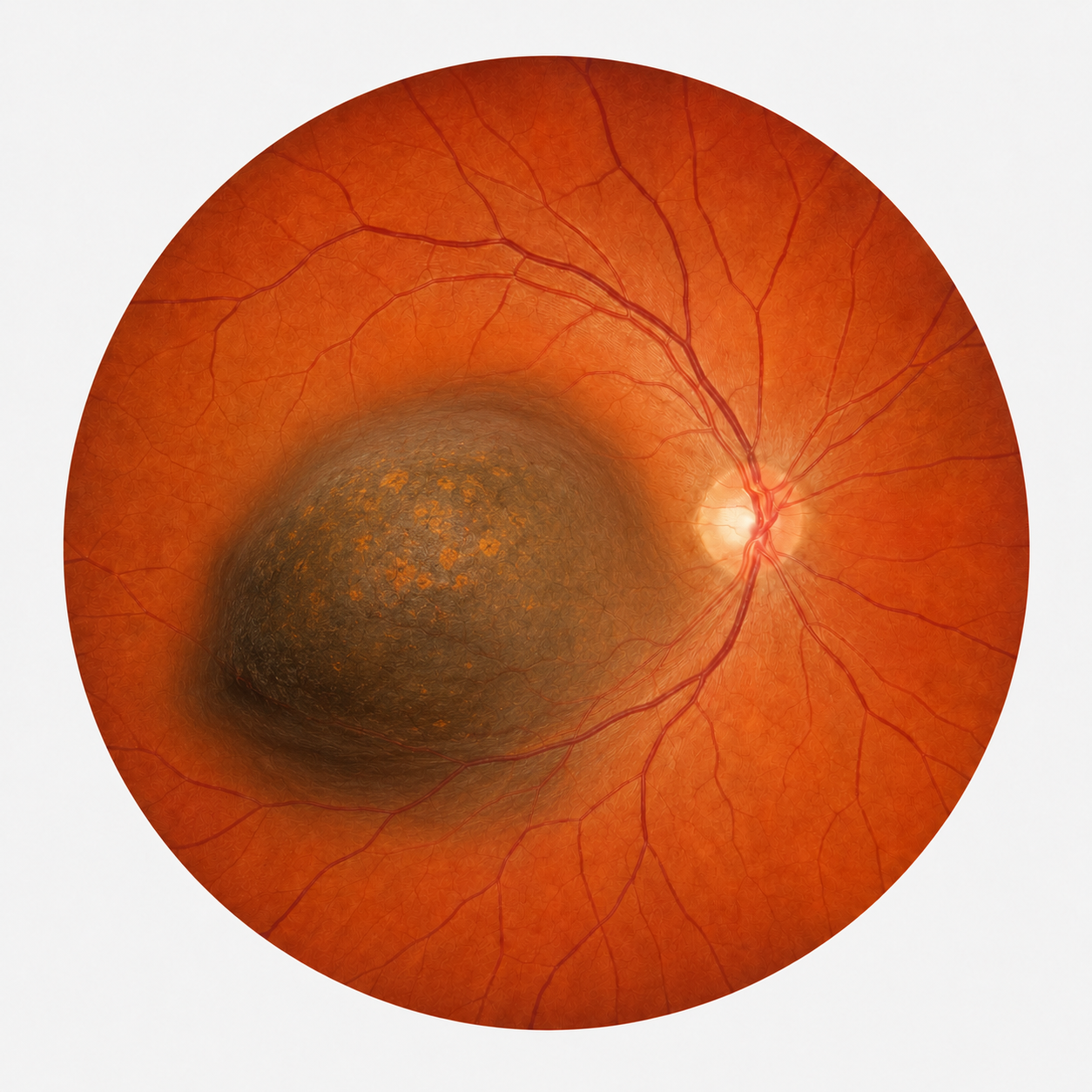
Normal Retina vs. Choroidal Melanoma
Choroidal melanoma may appear as a dark pigmented lesion beneath the retina. A retina specialist can evaluate the size, appearance, and growth of suspicious lesions.
Symptoms
Symptoms of Choroidal Melanoma
Most patients exhibit no symptoms of choroidal melanoma and it is only discovered through routine eye examinations. If symptoms do occur, the patient might experience the following:
Causes
Causes of Choroidal Melanoma
The exact cause of choroidal melanoma is not known, however, there is a strong association with exposure to ultraviolet (UV) light. Also, people with light eye color, of Caucasian descent, and older in age are at greater risk. Some skin disorders, such as dysplastic nevus syndrome or abnormal skin pigmentation of the face, are also at a greater risk for choroid melanoma.
Evaluation
Early Detection Matters
Because many patients do not notice symptoms early, routine dilated eye examinations are important. A retina specialist can evaluate suspicious pigmented lesions in the back of the eye and monitor for growth or changes over time.
Treatment depends on the size and location of the tumor.
The primary goal is to kill the tumor and decrease the chance of it spreading to the body. The second goal is to save as much vision as possible.
Treatment
Treating Choroidal Melanoma
The treatment of choroidal melanoma will depend largely on the size and location in the eye. The primary goal is to kill the tumor and decrease the chance of it spreading to the body. The second goal is to save as much vision as possible. A small choroidal melanoma might not need surgery right away, but requires close observation of the growth.
Radioactive Plaque Treatments
Radioactive Plaque Treatments
A patient with a medium or large-sized choroidal melanoma is generally treated with radiation therapy or laser treatment. Ophthalmic plaque radiation therapy is the most common therapy used, with a proven track record, that limits the need to remove part of the eye. The radiation will cause some decrease in vision over time, but rarely do patients lose all vision from this treatment.
Dr. Mason has been performing radioactive plaque treatments of melanoma since 1994. The radiation plaque treatment has a 90% chance of killing the tumor, with only a 10% chance of tumor recurrence. Laser treatment in the office typically has a 70% chance of killing small melanomas, but is not very effective for medium-sized and larger melanomas.
Schedule an Appointment
Call to schedule an appointment today.
Our retina specialists provide evaluation and treatment for diseases affecting the retina, macula, vitreous, and choroid.